Unity Health – St. Michael’s Hospital is seeking a solution to detect and assess cerebrovascular pathological segments in venous and carotid segments to confirm appropriate therapeutic intervention.
Unity Health – St. Michael’s Hospital is posting this Call for Innovation to seek out qualified Canadian companies who can meet the desired outcomes. Unity Health – St. Michael’s Hospital and CAN Health reserves the right to not move forward with this project at its full discretion and in particular if there are no qualified Canadian companies that can reasonably meet the desired outcomes.
To qualify for a CAN Health project, the company must have its headquarter in Canada and/or the majority (>50%) of the company owned by Canadians and/or significant economic impact to Canada including a high Canadian job creation potential, >70% of contract value to Canada (for distributors of a non-Canadian solution), independent autonomy over business operations and product development (for subsidiaries, affiliates or distributors), current presence (office(s) and client(s)) and can benefit from the CAN Health Network. Priority will be given to companies that meet all eligibility criteria.
For more information on the Call for Innovation process and the commercialization projects funded by CAN Health Network, please refer to the FAQ page on the CAN Health Network website: https://canhealthnetwork.ca/faq/
Problem Statement: Stroke and neuro centers across Canada are grappling with the intricate challenge of accurately diagnosing and effectively treating cerebral pathologies because of the lack of secondary imaging capabilities.
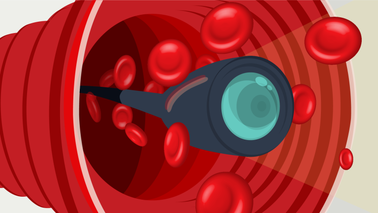
Objectives: Unity Health is focused on improving patient outcomes by deploying an innovative intravascular imaging system that features neuro-angioscopy. This technology offers direct, real-time, endoluminal, and color imaging, tailored for neurovascular indications.
Essential Requirements (all sections are mandatory)
The proposed solution must:
Intended Use:
- The intended for use of the solution must be use for directly visualizing the interiors of blood vessels within both peripheral and cerebral vasculature.
Image Clarity and Resolution:
- The ability of the solution to produce clear and high-resolution images that allow for the differentiation between acute and chronic occlusions.
Size and Configuration:
- Digital Segment OD – <0.72mm
- Proximal Segment OD – <0.80mm
- Working Length – >155cm
- Tip Characteristics – Straight
- Field of View (FOV) – 55
- Direction of View (DOV) – 0
- Maximum Insertion Portion Width – <0.80mm
Equipment Compatibility:
- Must be compatible with Karl Storz, Olympus, Stryker and other standard off-the-shelf light sources and endoscopy platforms
Hydrophilic Coating:
- The solution must posses a minimum hydrophilic coating length, from the distal tip, of 110cm
Flexibility and Trackability:
- The solution must provide exceptional flexibility and trackability to navigate challenging and tortuous neurovascular pathways while maintaining precise control during catheter advancement.
Radiopacity and Visualization:
- Exceptional radiopacity is paramount for accurate placement and real-time visualization of the catheter and associated devices during fluoroscopic and angiographic procedures in the neurovascular system. The solution must contain a minimum of 1 radiopaque marker at the distal tip and the total length must be radiopaque.
Packaging and Sterilization:
- The solution must be individually packaged, sterile, and ready for immediate use upon opening. The packaging should maintain product sterility until use. Devices that require reprocessing and sterilization will not be considered.
Quality Assurance:
- The chosen supplier should have a robust quality assurance and control system in place, including regular quality audits and reporting. They must be ISO13485/MDASAP certified.
Device Durability and Maintenance:
- The solution must be a single-use device that does not require additional maintenance and upkeep.
Quantities and Delivery:
- The EDGE anticipates a recurring need for a solution to support our ongoing neurointerventional stroke treatments. The supplier must be capable of providing a sufficient quantity of solution on a regular basis and adhere to agreed-upon delivery schedules to meet the urgent demands of stroke cases.
Contraindications:
The solution must NOT be contraindicated for any of the following:
- Acute renal failure
- Presence of a large thrombus
- Major abnormalities in the coagulation system
- Patients diagnosed with spasm
- Patients disqualified for Coronary Artery Bypass Grafting (CABG)
- Patients disqualified for Percutaneous Transluminal Coronary Angioplasty (PTCA)
- Severe hemodynamic instability or shock
- Total occlusion
Cerebrovascular disease is a heterogeneous disorder comprising several distinct pathologies, including transient ischemic attack, different types of strokes (ischemic stroke, intracerebral hemorrhage, subarachnoid hemorrhage), and various intracranial vascular disorders like vascular malformations and unruptured aneurysms (Sharma, 2017). Cerebrovascular diseases, as highlighted by Sharma (2017), Khaku (2023), and Tong X (2019), present a significant global health challenge, leading to high mortality rates and pervasive long-term disabilities. This places an enormous burden on the Canadian Healthcare System.
Stroke, a significant aspect of cerebrovascular diseases, is a leading cause of long-term disability in adults and ranks as the fifth leading cause of death worldwide. Despite a recent decrease in stroke incidence, the associated morbidity has risen substantially. Globally, it’s estimated that at least 5 million people die from strokes, with millions more living with disabilities (Khaku, 2023).
The management of these diseases is further complicated by the limitations of current diagnostic and therapeutic approaches. These statistics emphasize the critical need for improvements in diagnostic and therapeutic strategies (Tong X, 2019). Intravascular visualization through angiograms (fluoroscopy, 3D Angiogram, Cone Beam CT) remains the core assessment for diagnosis, treatment, and monitoring of cerebrovascular disorders.
The limitations of current imaging techniques in accurately visualizing intracranial vascular diseases are significant. These methods often struggle to comprehensively capture the complexities of diseases within intracranial vessels, highlighting the necessity for more advanced imaging tools. In response to this challenge, angioscopy is emerging as a complementary tool. Its integration is aimed at enhancing diagnostic capabilities, particularly in complex scenarios. By adding angioscopy to the existing array of imaging modalities, there is a potential for substantially improved patient outcomes through a more thorough and detailed understanding of intracranial vascular conditions.